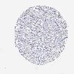
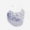
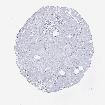
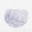
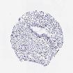
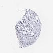
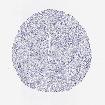
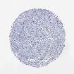
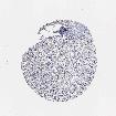
The primary data page shows the detailed antibody staining in 83 different cell types for each analyzed antibody
and in cases where a knowledge-based annotated protein expression profile is available, this
result is displayed in an additional column to the right. The level of expression is also given by a blue-scale
color-coding (described by the scale in the box to the right). More detailed information can be found under
Assays & annotation. The antibodies analyzed are listed in the "Antibodies in assay"-field.
The tissues and cell types can be ordered by organ, cell type or alphabetically. The organ view displays the representative
tissues organized in tissue groups according to functional features or anatomical location. The cell type view displays the
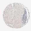
representative tissues organized in groups related to origin. The view allows the viewer to identify cell type specific
patterns of expression e.g. CD20 expression in hematopoietic cell types.
For each tissue and cell type, the antibody staining is given with the blue-scale color-coding (described by the scale
in the box to the right). Each available antibody is listed in a separate column and the antibody identifier is available
at the bottom of the tissue list.
The images and annotations can be accessed by clicking on the tissue name.
For histological reference, visit the
histological dictionary.