Stomach, lower
Proximal part (fundus-corpus)
The stomach lies in the upper part of the abdomen between the esophagus and duodenum which forms the most proximal portion of the small intestine. It mixes food with gastric enzymes and fluids, converting the contents to a semi fluid mass of partly digested food (termed chyme). The chyme is then slowly passed to the duodenum for further breakdown and absorption.
The stomach is a direct continuation of the esophagus and can be divided into different regions; the most proximal part is the cardia, followed by the fundus, corpus, antrum and pylorus. The fundus and corpus constitute about 80% of the stomach and differs from the antrum and pylorus both functionally and histologically.
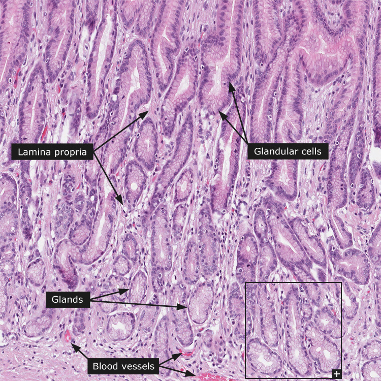
At the gastroesophageal junction the mucosa abruptly changes from stratified squamous epithelium to simple cuboidal. The mucosa is thick and lined with simple columnar epithelium. The surface epithelium invaginates into gastric pits into which the fundus glands open. The fundus glands are straight glands that extend from the lowest portion of the mucosa to their opening in the bottom of the gastric pits. The fundus glands stain darker compared to the gastric pits. In the stomach, the lamina muscularis mucosae consist of two layers of smooth muscle and can easily be recognized. Thesubmucosa is a thick layer of loose connective tissue with numerous blood and lymphatic vessels.
Villous folds separate the gastric pits and display a connective tissue core, which is part of the lamina propria mucosae. The gastric pits are lined by pale stained simple columnar epithelium that secrete mucous into the stomach lumen. This mucous protects the stomach wall from the acidic gastric contents. A mucinous globule is typically present in the apical portion of the cells. The nucleus is oval in shape.
The fundus glands have a narrow neck, a middle principal part and a lower base. In the neck portion, mucous neck cells are located. Their cytoplasm stain poorly with HE, but they can be recognized by their round nucleus. They secrete acidic mucous.
In the principle part, parietal (oxyntic) cells are located. They have strongly eosinophilic cytoplasm and a round, centrally located nucleus. They secrete hydrochloric acid and intrinsic factor. At the base of the glands, strongly basophilic cells are present. These are the chief cells that secrete large amount of pepsinogen, a proteolytic enzyme.
Distal part (antrum-pylorus)
The distal portion of the stomach, or the pylorus, connects the stomach to the duodenum. It has the same general structure as the fundus and body of the stomach with epithelial lined villous folds that invaginate into gastric pits. The cells lining the villous folds are surface mucus cells that produce alkaline mucus to protect the gastric mucosa from the acidic content of the stomach. At the bottom of the gastric pits the pyloric glands open. Instead of being simple tubular glands as the fundus glands, they are branched tubular glands. The cells of the pyloric glands are almost exclusively mucous secreting, resembling the mucous neck cells of the fundus glands. Within the pyloric glands gastrin producing enteroendocrine cells are also present.
The general structure of all parts of the GI-tract is
1) tunica serosa /adventitia - Loose connective tissue with elastic and collagen fibers, nerves and vessels, covered by a single layer of flat mesothelial cells. Where there is no mesothelial cover the outermost layer is called adventitia.
2) tela subserosa - thin layer of loose connective tissue separating the serosa and muscle layer.
3) tunica muscularis - which for most parts is composed of an inner circular and outer longitudinal smooth muscle layer. Between the muscle fibers the myenteric plexus of Auerbach can be identified.
4) tela submucosa - a thick layer of loose connective tissue with numerous of blood and lymphatic vessels. Here is where the ganglion cells of the submucosal plexus of Meissner might be seen.
5) tunica mucosa - the innermost layer that comes in contact with the gastrointestinal content. It has secretory and absorptive function. The mucosa consists of the innermost epithelium that forms surface cells and glands, embedded in the lamina propria containing mainly of loose connective tissue with small blood vessels and immune cells. A thin layer of smooth muscle, lamina muscularis mucosae, demarcates the division of the mucosa and submucosa.
Cancer: Stomach cancer